LGA
Sequence Independent Analysis (LGA)
Frame of reference: Cat.Q2246_545_244.5wLII_11212_65
Total number of 3D structures: 16
LGA calculations using distance cutoff DIST: 4.0 A
Residues superimposed below 2.00 A: GREEN
Residues superimposed below 4.00 A: YELLOW
Residues superimposed below 6.00 A: ORANGE
Residues superimposed below 8.00 A: BROWN
Residues superimposed above 8.00 A or not aligned: RED
Terminal residues not aligned: GREY
Structure Deviation Summary
Calculations based on one final LGA superposition
(Bar representation of 3D plots, TEXT)
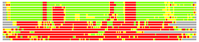
Structures ordered by LGA_S - score
| Structure |
NS |
NT |
N(dist=4.0) |
RMSD(N) |
Seq_ID(N) |
LGA_S |
LGA_Q |
PLOTS |
| 1ytm_A |
517 |
193 |
168 |
1.09 |
16.07 |
84.176 |
14.064 |
T P |
| 1ii2_A |
522 |
193 |
165 |
1.37 |
11.52 |
81.920 |
11.207 |
T P |
| 2py7_X |
535 |
193 |
160 |
1.44 |
13.75 |
79.327 |
10.377 |
T P |
| 2olr_A |
535 |
193 |
158 |
1.33 |
13.92 |
78.588 |
11.017 |
T P |
| 1ayl_A |
532 |
193 |
158 |
1.33 |
13.92 |
77.816 |
11.072 |
T P |
| 1ylh_A |
524 |
193 |
157 |
1.64 |
13.38 |
74.434 |
9.031 |
T P |
| 1j3b_B |
518 |
193 |
155 |
1.72 |
14.84 |
73.884 |
8.504 |
T P |
| 1oen_A |
524 |
193 |
155 |
1.71 |
14.19 |
73.714 |
8.547 |
T P |
| 1yip_A |
311 |
193 |
45 |
2.53 |
2.22 |
16.558 |
1.712 |
T P |
| 1yjl_A |
285 |
193 |
44 |
2.56 |
2.27 |
15.656 |
1.656 |
T P |
| 1phm_A |
305 |
193 |
42 |
2.71 |
2.38 |
14.782 |
1.495 |
T P |
| 1sdw_A |
312 |
193 |
41 |
2.41 |
0.00 |
14.371 |
1.636 |
T P |
| 1l9x_B |
294 |
193 |
39 |
2.51 |
10.26 |
14.263 |
1.494 |
T P |
| 2iqc_A |
173 |
193 |
41 |
2.70 |
12.20 |
13.735 |
1.462 |
T P |
| 1yi9_A |
295 |
193 |
37 |
2.84 |
2.70 |
12.795 |
1.259 |
T P |
| 1yjk_A |
303 |
193 |
30 |
2.87 |
3.33 |
11.129 |
1.011 |
T P |
NS : Total number of residues in Structure (rotated structure)
NT : Total number of residues in TARGET (frame of reference)
N : Total number of residues superimposed under 4.0 Angstrom distance cutoff
RMSD : RMS deviation calculated on all N residues superimposed under 4.0 Angstrom distance cutoff
Seq_Id : Sequence Identity. Percent of identical residues from the total of N aligned.
LGA_S : Structure similarity score calculated by internal LGA procedure (see LGA paper for details)
LGA_Q : Score (how tight is the superposition) calculated by the formula: Q = 0.1*N/(0.1+RMSD)
PLOTS : T - Flat text file (output from LGA program, rotated structure)
PLOTS : P - Plot of superimposed structures (3D plot colored as bars)
Citing LGA:
Zemla A., "LGA - a Method for Finding 3D Similarities in Protein Structures",
Nucleic Acids Research, 2003, Vol. 31, No. 13, pp. 3370-3374.
[MEDLINE]