LGA
Sequence Independent Analysis (LGA)
Frame of reference: Cat.Q2246_545_346.5wLII_11276_148
Total number of 3D structures: 15
LGA calculations using distance cutoff DIST: 4.0 A
Residues superimposed below 2.00 A: GREEN
Residues superimposed below 4.00 A: YELLOW
Residues superimposed below 6.00 A: ORANGE
Residues superimposed below 8.00 A: BROWN
Residues superimposed above 8.00 A or not aligned: RED
Terminal residues not aligned: GREY
Structure Deviation Summary
Calculations based on one final LGA superposition
(Bar representation of 3D plots, TEXT)
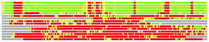
Structures ordered by LGA_S - score
| Structure |
NS |
NT |
N(dist=4.0) |
RMSD(N) |
Seq_ID(N) |
LGA_S |
LGA_Q |
PLOTS |
| 1thz_A |
590 |
279 |
242 |
1.23 |
11.57 |
84.264 |
18.238 |
T P |
| 1g8m_A |
590 |
279 |
242 |
1.25 |
11.57 |
84.217 |
17.966 |
T P |
| 1pkx_B |
589 |
279 |
242 |
1.32 |
12.81 |
83.503 |
16.993 |
T P |
| 1m9n_A |
590 |
279 |
240 |
1.16 |
11.67 |
83.459 |
19.041 |
T P |
| 1zcz_B |
454 |
279 |
200 |
1.68 |
13.50 |
61.630 |
11.235 |
T P |
| 1br2_A |
673 |
279 |
74 |
2.55 |
8.11 |
18.000 |
2.793 |
T P |
| 2aw5_B |
536 |
279 |
71 |
2.43 |
8.45 |
17.446 |
2.808 |
T P |
| 3b5v_A |
247 |
279 |
72 |
2.65 |
8.33 |
16.066 |
2.618 |
T P |
| 2qf7_A |
1075 |
279 |
60 |
2.74 |
6.67 |
14.643 |
2.109 |
T P |
| 2yv1_A |
268 |
279 |
57 |
2.52 |
8.77 |
14.396 |
2.175 |
T P |
| 2vqd_A |
447 |
279 |
55 |
2.66 |
12.73 |
13.661 |
1.992 |
T P |
| 1br1_A |
787 |
279 |
59 |
2.85 |
5.08 |
13.577 |
1.999 |
T P |
| 3fhl_C |
338 |
279 |
50 |
2.70 |
0.00 |
11.859 |
1.783 |
T P |
| 1g57_B |
209 |
279 |
49 |
2.67 |
8.16 |
11.813 |
1.767 |
T P |
| 1iez_A |
217 |
279 |
42 |
3.00 |
9.52 |
9.517 |
1.357 |
T P |
NS : Total number of residues in Structure (rotated structure)
NT : Total number of residues in TARGET (frame of reference)
N : Total number of residues superimposed under 4.0 Angstrom distance cutoff
RMSD : RMS deviation calculated on all N residues superimposed under 4.0 Angstrom distance cutoff
Seq_Id : Sequence Identity. Percent of identical residues from the total of N aligned.
LGA_S : Structure similarity score calculated by internal LGA procedure (see LGA paper for details)
LGA_Q : Score (how tight is the superposition) calculated by the formula: Q = 0.1*N/(0.1+RMSD)
PLOTS : T - Flat text file (output from LGA program, rotated structure)
PLOTS : P - Plot of superimposed structures (3D plot colored as bars)
Citing LGA:
Zemla A., "LGA - a Method for Finding 3D Similarities in Protein Structures",
Nucleic Acids Research, 2003, Vol. 31, No. 13, pp. 3370-3374.
[MEDLINE]