LGA
Sequence Independent Analysis (LGA)
Frame of reference: Cat.Q2246_545_473.5wLII_11346_51
Total number of 3D structures: 13
LGA calculations using distance cutoff DIST: 4.0 A
Residues superimposed below 2.00 A: GREEN
Residues superimposed below 4.00 A: YELLOW
Residues superimposed below 6.00 A: ORANGE
Residues superimposed below 8.00 A: BROWN
Residues superimposed above 8.00 A or not aligned: RED
Terminal residues not aligned: GREY
Structure Deviation Summary
Calculations based on one final LGA superposition
(Bar representation of 3D plots, TEXT)
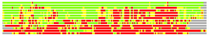
Structures ordered by LGA_S - score
| Structure |
NS |
NT |
N(dist=4.0) |
RMSD(N) |
Seq_ID(N) |
LGA_S |
LGA_Q |
PLOTS |
| 2gel_A |
218 |
214 |
209 |
0.45 |
22.49 |
97.305 |
38.012 |
T P |
| 1okj_B |
222 |
214 |
209 |
0.88 |
22.49 |
95.751 |
21.291 |
T P |
| 2a6a_A |
193 |
214 |
168 |
1.85 |
17.26 |
66.634 |
8.626 |
T P |
| 3eno_A |
329 |
214 |
156 |
2.16 |
18.59 |
55.868 |
6.891 |
T P |
| 3en9_A |
519 |
214 |
153 |
2.09 |
19.61 |
54.938 |
6.999 |
T P |
| 2vwb_A |
519 |
214 |
151 |
2.12 |
18.54 |
53.683 |
6.787 |
T P |
| 2ivn_A |
325 |
214 |
140 |
2.30 |
15.71 |
46.725 |
5.825 |
T P |
| 1hux_A |
259 |
214 |
119 |
2.49 |
7.56 |
37.859 |
4.601 |
T P |
| 2v7y_A |
504 |
214 |
127 |
2.48 |
4.72 |
37.709 |
4.920 |
T P |
| 1tel_B |
428 |
214 |
58 |
2.66 |
5.17 |
18.688 |
2.102 |
T P |
| 2dg1_B |
322 |
214 |
63 |
2.89 |
3.17 |
18.284 |
2.109 |
T P |
| 2gls_A |
468 |
214 |
50 |
2.43 |
4.00 |
17.151 |
1.974 |
T P |
| 2dso_C |
323 |
214 |
55 |
2.81 |
14.55 |
15.918 |
1.889 |
T P |
NS : Total number of residues in Structure (rotated structure)
NT : Total number of residues in TARGET (frame of reference)
N : Total number of residues superimposed under 4.0 Angstrom distance cutoff
RMSD : RMS deviation calculated on all N residues superimposed under 4.0 Angstrom distance cutoff
Seq_Id : Sequence Identity. Percent of identical residues from the total of N aligned.
LGA_S : Structure similarity score calculated by internal LGA procedure (see LGA paper for details)
LGA_Q : Score (how tight is the superposition) calculated by the formula: Q = 0.1*N/(0.1+RMSD)
PLOTS : T - Flat text file (output from LGA program, rotated structure)
PLOTS : P - Plot of superimposed structures (3D plot colored as bars)
Citing LGA:
Zemla A., "LGA - a Method for Finding 3D Similarities in Protein Structures",
Nucleic Acids Research, 2003, Vol. 31, No. 13, pp. 3370-3374.
[MEDLINE]