LGA
Sequence Independent Analysis (LGA)
Frame of reference: Cat.Q2246_545_528.5wLII_11390_17
Total number of 3D structures: 18
LGA calculations using distance cutoff DIST: 4.0 A
Residues superimposed below 2.00 A: GREEN
Residues superimposed below 4.00 A: YELLOW
Residues superimposed below 6.00 A: ORANGE
Residues superimposed below 8.00 A: BROWN
Residues superimposed above 8.00 A or not aligned: RED
Terminal residues not aligned: GREY
Structure Deviation Summary
Calculations based on one final LGA superposition
(Bar representation of 3D plots, TEXT)
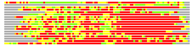
Structures ordered by LGA_S - score
| Structure |
NS |
NT |
N(dist=4.0) |
RMSD(N) |
Seq_ID(N) |
LGA_S |
LGA_Q |
PLOTS |
| 1qsa_A |
618 |
109 |
63 |
2.28 |
7.94 |
43.398 |
2.648 |
T P |
| 3epv_A |
109 |
109 |
39 |
2.26 |
5.13 |
30.106 |
1.650 |
T P |
| 3eie_A |
303 |
109 |
46 |
2.40 |
4.35 |
28.730 |
1.840 |
T P |
| 1f5n_A |
570 |
109 |
42 |
2.46 |
7.14 |
27.884 |
1.638 |
T P |
| 2ce7_C |
421 |
109 |
39 |
2.33 |
10.26 |
27.604 |
1.607 |
T P |
| 1qu7_A |
227 |
109 |
37 |
2.29 |
5.41 |
26.951 |
1.549 |
T P |
| 3eih_A |
319 |
109 |
46 |
2.71 |
4.35 |
26.486 |
1.639 |
T P |
| 2qp9_X |
288 |
109 |
40 |
2.94 |
2.50 |
25.232 |
1.314 |
T P |
| 1yqi_C |
708 |
109 |
41 |
2.68 |
4.88 |
24.931 |
1.475 |
T P |
| 2ch7_B |
307 |
109 |
35 |
2.36 |
2.86 |
24.792 |
1.425 |
T P |
| 3d8b_A |
281 |
109 |
37 |
2.53 |
10.81 |
24.473 |
1.404 |
T P |
| 3b9p_A |
268 |
109 |
38 |
2.75 |
10.53 |
23.767 |
1.332 |
T P |
| 2ch7_A |
309 |
109 |
35 |
2.55 |
8.57 |
23.597 |
1.319 |
T P |
| 1s3s_F |
441 |
109 |
39 |
3.04 |
2.56 |
23.382 |
1.243 |
T P |
| 2rko_A |
280 |
109 |
37 |
2.65 |
5.41 |
22.227 |
1.345 |
T P |
| 1ypw_A |
692 |
109 |
34 |
2.63 |
2.94 |
22.109 |
1.244 |
T P |
| 1r7r_A |
683 |
109 |
33 |
2.95 |
9.09 |
20.965 |
1.080 |
T P |
| 3cf2_A |
659 |
109 |
27 |
2.68 |
3.70 |
17.134 |
0.973 |
T P |
NS : Total number of residues in Structure (rotated structure)
NT : Total number of residues in TARGET (frame of reference)
N : Total number of residues superimposed under 4.0 Angstrom distance cutoff
RMSD : RMS deviation calculated on all N residues superimposed under 4.0 Angstrom distance cutoff
Seq_Id : Sequence Identity. Percent of identical residues from the total of N aligned.
LGA_S : Structure similarity score calculated by internal LGA procedure (see LGA paper for details)
LGA_Q : Score (how tight is the superposition) calculated by the formula: Q = 0.1*N/(0.1+RMSD)
PLOTS : T - Flat text file (output from LGA program, rotated structure)
PLOTS : P - Plot of superimposed structures (3D plot colored as bars)
Citing LGA:
Zemla A., "LGA - a Method for Finding 3D Similarities in Protein Structures",
Nucleic Acids Research, 2003, Vol. 31, No. 13, pp. 3370-3374.
[MEDLINE]