LGA
Sequence Independent Analysis (LGA)
Frame of reference: Cat.Q2246_545_76.5wLII_11067_10
Total number of 3D structures: 13
LGA calculations using distance cutoff DIST: 4.0 A
Residues superimposed below 2.00 A: GREEN
Residues superimposed below 4.00 A: YELLOW
Residues superimposed below 6.00 A: ORANGE
Residues superimposed below 8.00 A: BROWN
Residues superimposed above 8.00 A or not aligned: RED
Terminal residues not aligned: GREY
Structure Deviation Summary
Calculations based on one final LGA superposition
(Bar representation of 3D plots, TEXT)
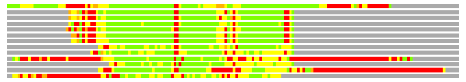
Structures ordered by LGA_S - score
| Structure |
NS |
NT |
N(dist=4.0) |
RMSD(N) |
Seq_ID(N) |
LGA_S |
LGA_Q |
PLOTS |
| 1qvr_A |
803 |
168 |
115 |
2.10 |
21.74 |
56.655 |
5.239 |
T P |
| 2iw5_A |
666 |
168 |
74 |
1.80 |
6.76 |
41.336 |
3.895 |
T P |
| 2h94_A |
647 |
168 |
74 |
1.86 |
6.76 |
41.016 |
3.772 |
T P |
| 2hko_A |
647 |
168 |
74 |
1.69 |
6.76 |
40.941 |
4.145 |
T P |
| 2v1d_A |
666 |
168 |
73 |
1.83 |
6.85 |
40.684 |
3.790 |
T P |
| 2dw4_A |
634 |
168 |
72 |
1.75 |
6.94 |
40.219 |
3.886 |
T P |
| 2z3y_A |
643 |
168 |
73 |
1.84 |
6.85 |
40.164 |
3.760 |
T P |
| 1qu7_A |
227 |
168 |
68 |
1.97 |
10.29 |
36.235 |
3.283 |
T P |
| 2ch7_B |
307 |
168 |
69 |
2.15 |
5.80 |
34.948 |
3.068 |
T P |
| 2b9m_Y |
365 |
168 |
72 |
2.27 |
12.50 |
33.662 |
3.034 |
T P |
| 2ch7_A |
309 |
168 |
65 |
2.13 |
7.69 |
32.773 |
2.921 |
T P |
| 1key_B |
221 |
168 |
63 |
2.20 |
9.52 |
30.644 |
2.737 |
T P |
| 2a01_A |
243 |
168 |
67 |
2.60 |
10.45 |
28.859 |
2.480 |
T P |
NS : Total number of residues in Structure (rotated structure)
NT : Total number of residues in TARGET (frame of reference)
N : Total number of residues superimposed under 4.0 Angstrom distance cutoff
RMSD : RMS deviation calculated on all N residues superimposed under 4.0 Angstrom distance cutoff
Seq_Id : Sequence Identity. Percent of identical residues from the total of N aligned.
LGA_S : Structure similarity score calculated by internal LGA procedure (see LGA paper for details)
LGA_Q : Score (how tight is the superposition) calculated by the formula: Q = 0.1*N/(0.1+RMSD)
PLOTS : T - Flat text file (output from LGA program, rotated structure)
PLOTS : P - Plot of superimposed structures (3D plot colored as bars)
Citing LGA:
Zemla A., "LGA - a Method for Finding 3D Similarities in Protein Structures",
Nucleic Acids Research, 2003, Vol. 31, No. 13, pp. 3370-3374.
[MEDLINE]